

Artificial Intelligence in Bioinformatics
Artificial Intelligence in Bioinformatics
1. Why AI and bioinformatics now converge
Biology became a data science before most laboratories noticed. A single RNA-sequencing run produces tens of millions of reads; a spatial experiment adds a coordinate to every measurement; a mass-spectrometry session generates spectra faster than any analyst can inspect them. The bottleneck moved long ago from generating data to making sense of it. Artificial intelligence (AI) is the set of methods that addresses that second problem, and over the past decade it has moved from a supporting role to the centre of the omics workflow.
Two developments explain the timing. First, omics platforms now produce structured, high-dimensional measurements at a scale that statistical learning thrives on. Second, the architectures behind modern AI, particularly the transformer, turned out to transfer remarkably well from text to biological sequences and to gene-expression matrices. The result is a field where a model pre-trained on tens of millions of cells, or hundreds of millions of protein sequences, can be adapted to a specific question with comparatively little new data.
This article is written for a mixed readership. It is didactic enough to follow without a machine-learning background, and precise enough to be useful to bioinformaticians and regulatory scientists. It covers the core families of AI methods, their concrete use in transcriptomics, proteomics and integrative omics, and the practical advantages they bring to toxicology and cosmetic safety assessment, where the regulatory pressure to replace animal testing has made computational evidence indispensable.
2. A short, honest primer on the AI toolbox
It helps to separate three terms that are often used interchangeably but mean different things in practice.
2.1 Machine learning
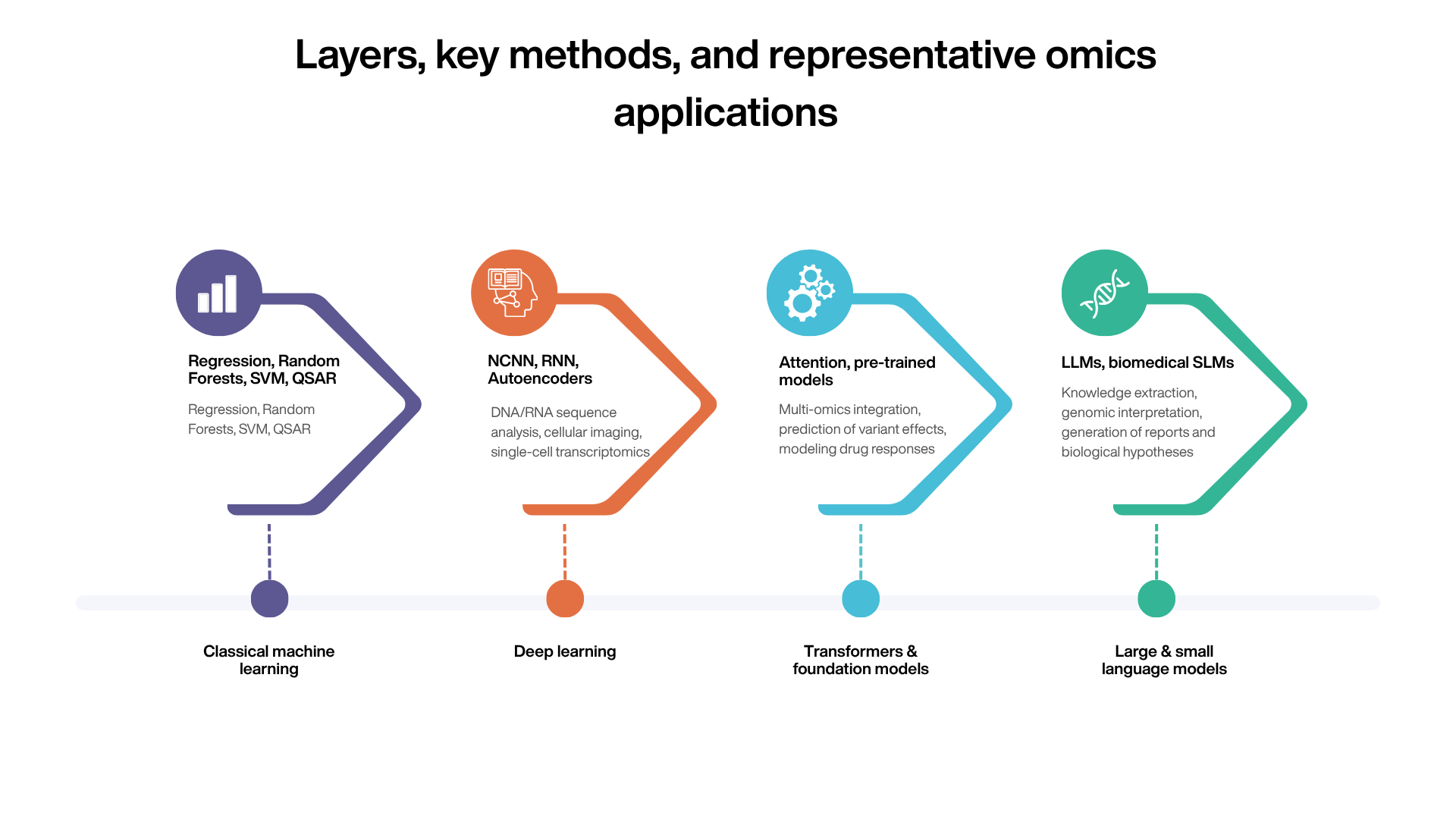
Machine learning (ML) covers algorithms that learn a mapping from inputs to outputs from examples, rather than from hand-written rules. In omics, the classical workhorses are penalised regression (LASSO, elastic net), random forests, gradient boosting and support vector machines. These methods remain competitive, and frequently superior, on tabular problems with modest sample sizes: biomarker selection, sample classification, dose-response modelling. They are interpretable, fast to train, and Wella understood statistically. Their main limitation is that they depend on features the analyst defines in advance, so a large part of the work happens before the model is trained.
2.2 Deep learning
Deep learning uses neural networks with many layers that learn their own representations of the data. Convolutional networks read local patterns in sequences and images; recurrent networks and, more recently, transformers handle ordered data such as nucleotide or amino-acid chains; autoencoders compress high-dimensional measurements into compact latent spaces useful for denoising and integration. Deep learning shines when data are abundant and the relevant features are not obvious in advance. It is more demanding in data and compute, and harder to interpret, which matters a great deal in a regulatory setting.
2.3 Foundation models, LLMs and SLMs
A foundation model is a large neural network pre-trained on a broad corpus using self-supervision, so that no manual labels are required, and then adapted to many downstream tasks. Large language models (LLMs) are foundation models trained on text. Small language models (SLMs) share the same architecture but with far fewer parameters, which makes them cheaper to run, easier to deploy on local infrastructure, and easier to specialise on a narrow domain. The key idea that carried over to biology is that a sequence of genes in a cell, or of amino acids in a protein, can be treated like a sentence: the model learns the grammar of biology by predicting masked or next tokens, and the representations it builds turn out to capture meaningful structure.
3. AI in transcriptomics
Transcriptomics measures which genes are active, and how strongly, across conditions, tissues or individual cells. The data are noisy, sparse (especially at single-cell resolution) and very high-dimensional. This combination is exactly where modern AI has made the clearest difference.
3.1 From bulk to single cell
In bulk RNA-seq, machine learning has long been used for sample classification, deconvolution of cell-type proportions, and signature discovery. The shift to single-cell and spatial transcriptomics raised the difficulty: millions of cells, dropout noise, and the need to align datasets generated by different laboratories and protocols. Deep generative models, in particular variational autoencoders, became standard tools for denoising, batch correction and dimensionality reduction, because they learn a probabilistic latent representation of each cell rather than relying on fixed features.
3.2 Single-cell foundation models
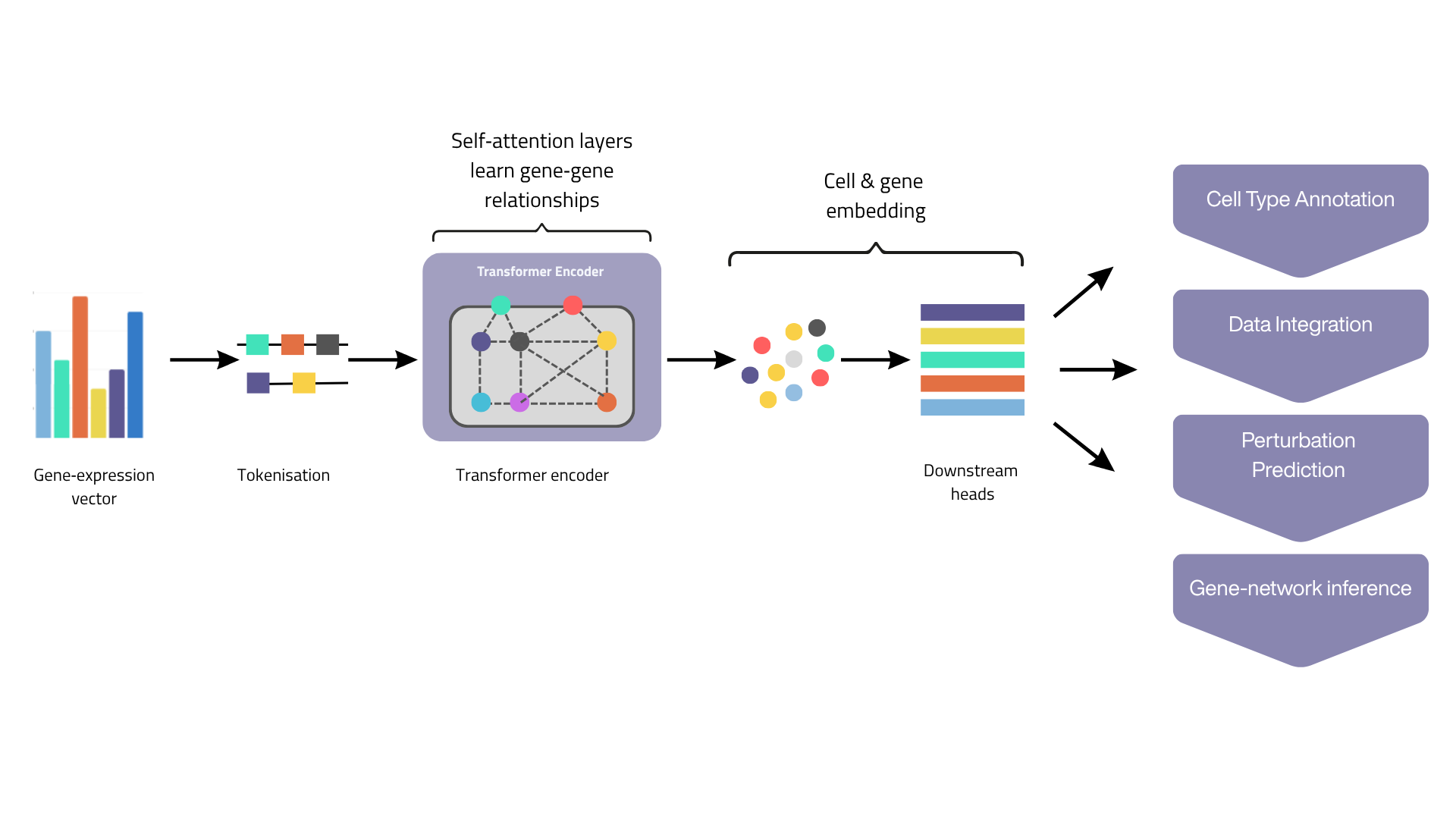
The most striking recent development is the single-cell foundation model. scGPT was pre-trained with a generative transformer on more than 33 million cells and can be fine-tuned for cell-type annotation, batch integration, gene-network inference and perturbation prediction[1]. Geneformer demonstrated that transfer learning on a large transcriptomic corpus enables predictions in network biology, including the in silico identification of candidate therapeutic targets[2]. scFoundation scaled this approach to a large-parameter model trained across tens of millions of cells[3]. The shared principle is simple to state: tokenise the expression profile of a cell, pre-train the model to reconstruct masked values, and reuse the learned embeddings for many tasks with little labelled data.
It is worth being candid about the limits. Independent benchmarks have shown that these models do not uniformly beat well-tuned classical pipelines; on some cell-type classification tasks a carefully applied principal component analysis or a method such as Seurat remains a strong baseline, and performance is uneven across models. The honest position is that single-cell foundation models are powerful, especially in low-data or noisy regimes, but they are tools to be benchmarked against simpler approaches rather than assumed to be superior.
4. AI in proteomics
Proteins are closer to phenotype than transcripts, and proteomics has its own analytical challenges: identifying peptides from fragmentation spectra, quantifying them reproducibly across runs, and understanding three-dimensional structure and interactions. AI has reshaped all three.
4.1 Structure prediction
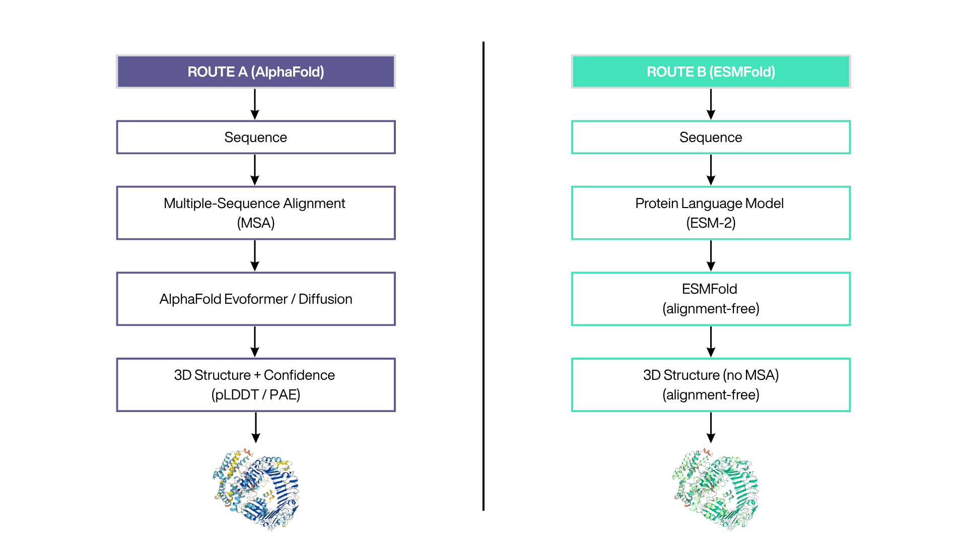
AlphaFold2 changed structural biology by predicting protein structures from sequence at an accuracy approaching experimental methods, a result confirmed in the blind CASP14 assessment[4]. AlphaFold3 extended the approach to complexes that include nucleic acids, small molecules, ions and modified residues within a single diffusion-based framework, with substantial gains for protein-ligand and protein-nucleic-acid interactions[5]. In parallel, protein language models took a different route: ESM-2, trained on hundreds of millions of sequences, learns structural information directly from sequence statistics, and its companion ESMFold predicts structure without a multiple-sequence alignment, fast enough to fold hundreds of millions of metagenomic proteins[6]. For applied work this means that a structural hypothesis for almost any protein of interest is now a starting point rather than a multi-year project.
4.2 Mass-spectrometry data analysis
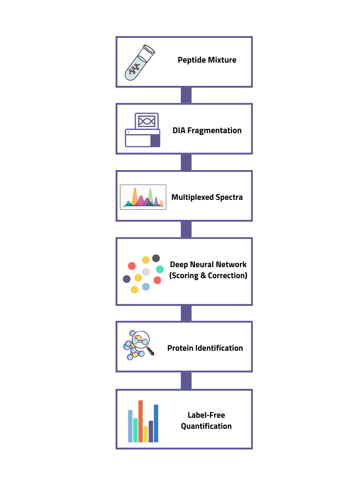
On the analytical side, deep learning has become standard in data-independent acquisition (DIA) proteomics. DIA-NN uses neural networks and a new signal-correction strategy to identify and quantify peptides reproducibly, enabling deep proteome coverage with fast chromatography and high-throughput study designs[7]. Related models predict fragment-ion intensities and retention times in silico, which improves spectral-library generation and peptide identification confidence. The practical consequence is more proteins quantified per sample, fewer missing values across large series, and workflows that scale to the sample numbers a service laboratory actually handles.
5. AI for integrative, multi-omics analysis
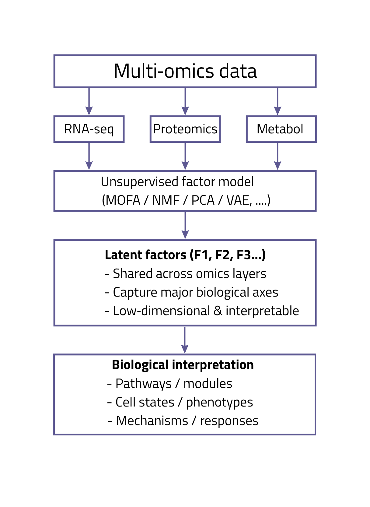
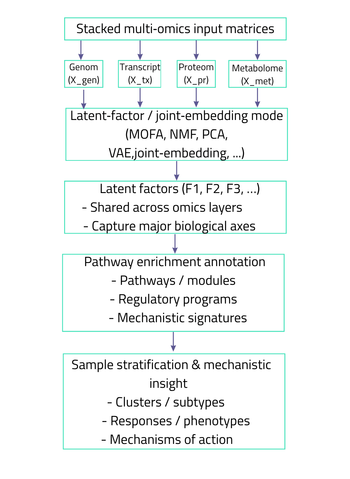
A single omics layer rarely tells the whole story. Genomics points to what could happen, transcriptomics to what is being transcribed, proteomics to what is active, and metabolomics to the biochemical consequences. Integrating these layers is a statistical problem of heterogeneous data types, different scales and many missing values.
Latent-variable models address this directly. Multi-Omics Factor Analysis (MOFA) generalises principal component analysis to several data matrices measured on the same samples, inferring interpretable factors that capture the main shared and modality-specific sources of variation[8]. Supervised and multivariate frameworks such as mixOmics and its DIABLO method select discriminant features across omics blocks and are widely used in applied biology[9].
More recent deep-learning approaches, built on autoencoders and attention, learn joint embeddings that can impute missing modalities and combine bulk with single-cell data. For interpretation, the field is moving toward methods that read out perturbed pathways and regulatory activity from these integrated representations, which is where the value lies for mechanistic and safety questions.
Language models contribute in two distinct ways, and conflating them causes most of the confusion around AI in biology.
6.1 Sequences as language
The first contribution treats biological sequences as text. Genomic language models such as the Nucleotide Transformer, pre-trained on genomes from many species, learn representations of DNA that transfer to tasks like predicting regulatory elements and the effects of variants[10]. Models such as Evo extend this idea across DNA, RNA and protein from the molecular to the genome scale, and can generate as well as predict sequences[11]. These are not chatbots; they are foundation models whose 'language' happens to be nucleotides or amino acids, and they sit upstream in analytical pipelines.
6.2 LLMs and SLMs as laboratory assistants
The second contribution is the familiar text-based LLM, used to read the literature, draft and debug analysis code, summarise results, annotate datasets, and orchestrate multi-step workflows as agents. For a service laboratory this is where day-to-day productivity gains are largest. Small language models matter here for a specific reason: a compact model fine-tuned on an organisation's own documents and run on local infrastructure keeps confidential client data in-house, costs less per query, and can be specialised to a narrow domain such as regulatory toxicology. Retrieval-augmented generation, which grounds a model's answers in a curated document base, is the standard way to reduce fabricated statements and keep outputs traceable to sources.
The caveats are real and should be stated plainly. Text LLMs can produce fluent but incorrect statements, they do not natively cite primary evidence, and they should not be treated as a source of biological truth. Used as assistants with a human expert in the loop and with outputs grounded in verifiable sources, they are valuable. Used as oracles, they are a liability.
7. Applications in toxicology
Toxicology has shifted from an observational discipline based on apical endpoints in animals toward a data-rich, mechanism-based science. This is the framework of New Approach Methodologies (NAMs): in silico, in chemico and in vitro methods that inform risk assessment without animal testing. AI is the connective tissue that makes NAM data usable at scale[12].
7.1 Predicting hazard from structure and bioactivity
The oldest computational toxicology methods are quantitative structure-activity relationships (QSAR), which relate molecular descriptors to toxicity. Deep learning extended this substantially: the DeepTox approach, trained on thousands of compounds in the Tox21 programme, matched or exceeded conventional methods at predicting toxicity endpoints[13]. Graph neural networks now represent molecules directly as graphs of atoms and bonds, and multi-task models predict several mechanistically related endpoints at once, which improves performance for endpoints with few active examples.
7.2 Reading mechanism from omics
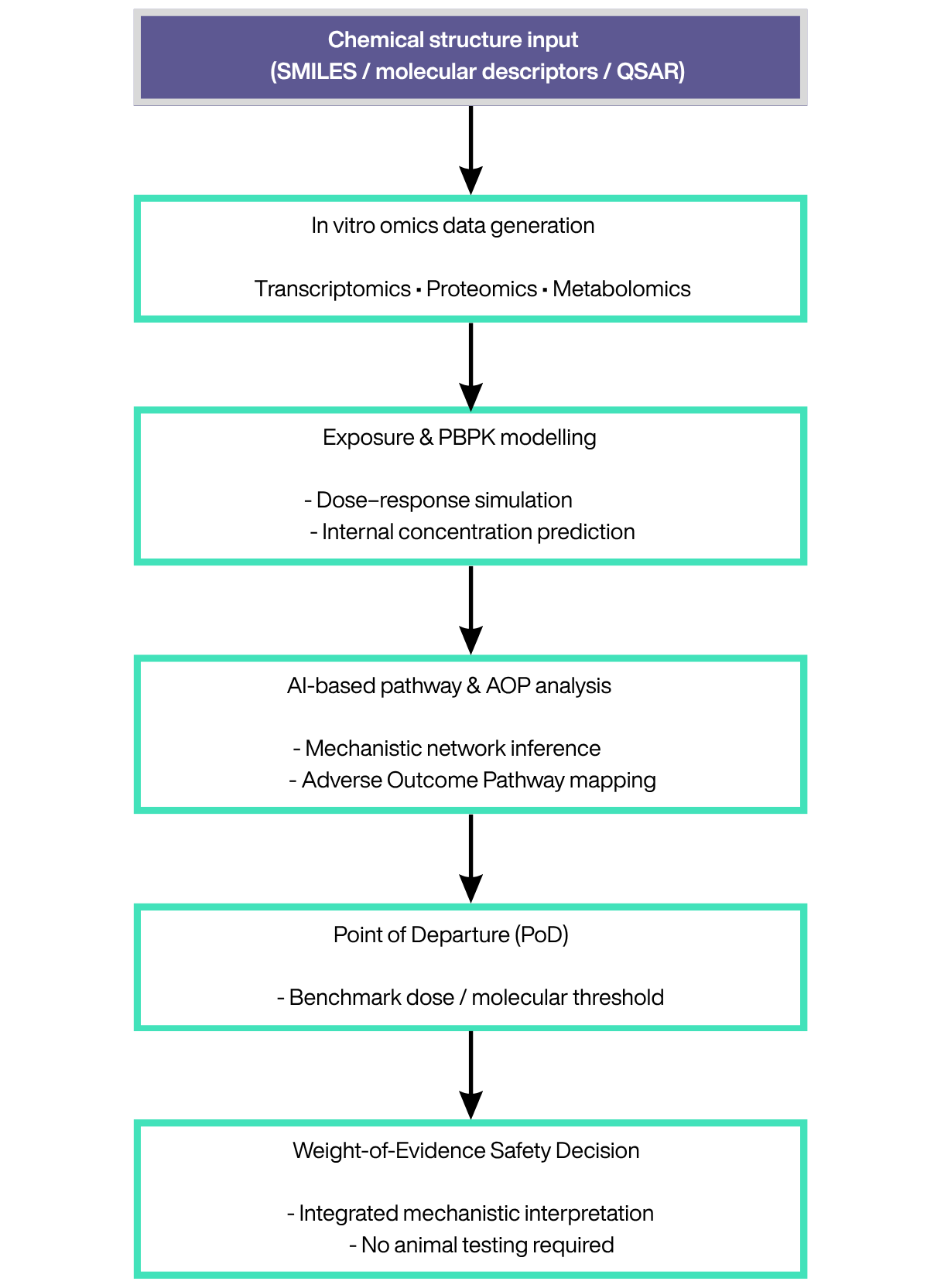
Where AI adds the most distinctive value is in interpreting omics responses to chemical exposure. Transcriptomic and proteomic signatures from cells exposed to a test substance can be clustered, mapped to perturbed pathways, and connected to adverse outcome pathways. Machine learning supports dimensionality reduction, exposure-signature clustering and the inference of points of departure for risk assessment. In image-based high-content screening, deep learning automates segmentation and detects subtle phenotypic changes that are hard to quantify by hand. The advantages are concrete: human-relevant mechanistic evidence, higher throughput, lower cost, and a path away from animal models.
The constraint is regulatory acceptance. Models used in safety decisions must be transparent, validated and reproducible, and the lack of mechanistic interpretability in some deep models, together with the need for large high-quality training data, remains the central obstacle to their formal use. This is why interpretability and rigorous benchmarking are not optional extras in this domain.
8. Applications in cosmetic safety assessment
Cosmetics is the field where the regulatory and scientific pressures meet most sharply. Under Regulation (EC) No 1223/2009, the European Union prohibits animal testing for cosmetic ingredients and finished products, with a full ban on all toxicological endpoints in force since March 2013[15]. Validated non-animal methods exist for local endpoints such as skin and eye irritation, skin sensitisation and phototoxicity, but systemic and repeated-dose toxicity remain difficult. The response is Next Generation Risk Assessment (NGRA): an exposure-led, hypothesis-driven framework that combines NAMs in a weight-of-evidence approach.
8.1 What AI contributes
AI enters NGRA at several points. In silico models predict physicochemical properties, skin penetration and metabolism, feeding physiologically based pharmacokinetic (PBPK) models that estimate internal exposure. Omics readouts from human cell systems are interpreted with machine learning to identify perturbed pathways and derive points of departure. An ab initio NGRA case study on the fragrance ingredient benzyl salicylate illustrates the approach: combining skin-absorption assays, hepatocyte metabolism, PBPK modelling, transcriptomics in human cell lines and cell-stress assays, the non-animal assessment reached the same safety conclusion as the traditional animal-data approach for a leave-on facial cream[14].
For skin science specifically, the combination is powerful. Transcriptomic and proteomic profiling of reconstructed human epidermis or skin models, analysed with the integrative methods described above, characterises a human-relevant response to an ingredient. AI turns that response into interpretable, decision-ready evidence: which pathways move, at what concentration, and how that compares with a benchmark of safe exposure. This is the core of animal-free safety substantiation, and it is where omics expertise and computational rigour together create defensible conclusions.
8.2 Beyond safety: efficacy and claims
The same toolbox supports efficacy substantiation and claims. Omics signatures can demonstrate that an active ingredient modulates a biological process of interest, for example pathways linked to barrier function, pigmentation or ageing, and machine learning helps separate genuine effects from noise across donors and conditions. Under the EU regime governing cosmetic claims, evidence of this quality is increasingly expected, and a defensible, mechanism-anchored dataset is a commercial as well as a scientific asset.
9. What to keep in mind
AI in omics is genuinely transformative, and it is also frequently oversold. A few principles keep its use honest:
• Benchmark against simple baselines. A foundation model that does not beat a well-tuned classical method on your task is not worth its complexity.
• Data quality dominates. Models inherit the biases of their training data, which over-represent well-studied organisms, tissues and conditions; conclusions outside that distribution deserve caution.
• Interpretability is a requirement, not a luxury, wherever a result feeds a safety or regulatory decision.
• Keep an expert in the loop. Language models accelerate work but do not replace scientific judgement, and ungrounded outputs should never be reported as fact.
• Match the tool to the constraint. Small, locally hosted models can be the right answer when confidentiality, cost and traceability matter more than raw scale.
10. Conclusion
Artificial intelligence has become part of the standard apparatus of bioinformatics, not a separate speciality. In transcriptomics it denoises, integrates and annotates at single-cell resolution. In proteomics it predicts structure from sequence and extracts deeper, more reproducible quantification from mass spectra. Across omics it integrates heterogeneous layers into interpretable factors. And in toxicology and cosmetics it converts mechanistic, human-relevant data into the kind of evidence that animal-free risk assessment now requires.
The value is not in the model alone but in the pairing of the right method with deep domain knowledge and a clear question. That pairing, omics expertise, regulatory understanding and computational rigour, is exactly where careful bioinformatics work earns its place. At SciLicium, this is the work we do every day.
References
1. Cui H, Wang C, Maan H, et al. scGPT: toward building a foundation model for single-cell multi-omics using generative AI. Nature Methods 21, 1470–1480 (2024). https://doi.org/10.1038/s41592-024-02201-0
2. Theodoris CV, Xiao L, Chopra A, et al. Transfer learning enables predictions in network biology. Nature 618, 616–624 (2023). https://doi.org/10.1038/s41586-023-06139-9
3. Hao M, Gong J, Zeng X, et al. Large-scale foundation model on single-cell transcriptomics. Nature Methods 21, 1481–1491 (2024). https://doi.org/10.1038/s41592-024-02305-7
4. Jumper J, Evans R, Pritzel A, et al. Highly accurate protein structure prediction with AlphaFold. Nature 596, 583–589 (2021). https://doi.org/10.1038/s41586-021-03819-2
5. Abramson J, Adler J, Dunger J, et al. Accurate structure prediction of biomolecular interactions with AlphaFold 3. Nature 630, 493–500 (2024). https://doi.org/10.1038/s41586-024-07487-w
6. Lin Z, Akin H, Rao R, et al. Evolutionary-scale prediction of atomic-level protein structure with a language model. Science 379, 1123–1130 (2023). https://doi.org/10.1126/science.ade2574
7. Demichev V, Messner CB, Vernardis SI, Lilley KS, Ralser M. DIA-NN: neural networks and interference correction enable deep proteome coverage in high throughput. Nature Methods 17, 41–44 (2020). https://doi.org/10.1038/s41592-019-0638-x
8. Argelaguet R, Velten B, Arnol D, et al. Multi-Omics Factor Analysis — a framework for unsupervised integration of multi-omics data sets. Molecular Systems Biology 14, e8124 (2018). https://doi.org/10.15252/msb.20178124
9. Rohart F, Gautier B, Singh A, Lê Cao K-A. mixOmics: An R package for 'omics feature selection and multiple data integration. PLoS Computational Biology 13, e1005752 (2017). https://doi.org/10.1371/journal.pcbi.1005752
10. Dalla-Torre H, Gonzalez L, Mendoza-Revilla J, et al. Nucleotide Transformer: building and evaluating robust foundation models for human genomics. Nature Methods 22, 287–299 (2025). https://doi.org/10.1038/s41592-024-02523-z
11. Nguyen E, Poli M, Durrant MG, et al. Sequence modeling and design from molecular to genome scale with Evo. Science 386, eado9336 (2024). https://doi.org/10.1126/science.ado9336
12. Hartung T. ToxAIcology — the evolving role of artificial intelligence in advancing toxicology and modernizing regulatory science. ALTEX 40, 559–570 (2023). https://doi.org/10.14573/altex.2309191
13. Mayr A, Klambauer G, Unterthiner T, Hochreiter S. DeepTox: Toxicity Prediction using Deep Learning. Frontiers in Environmental Science 3, 80 (2016). https://doi.org/10.3389/fenvs.2015.00080
14. Ebmeyer J, Najjar A, Lange D, et al. Next generation risk assessment: an ab initio case study to assess the systemic safety of the cosmetic ingredient, benzyl salicylate, after dermal exposure. Frontiers in Pharmacology 15, 1345992 (2024). https://doi.org/10.3389/fphar.2024.1345992
15. Regulation (EC) No 1223/2009 of the European Parliament and of the Council of 30 November 2009 on cosmetic products. Official Journal of the European Union (consolidated text). https://eur-lex.europa.eu/legal-content/EN/TXT/?uri=CELEX:02009R1223-20240501